CARDIAC SCANNING MADE EASY
High performance in image quality and speed must go hand in hand with efficient workflow. The Aquilion RXL has specifically addressed workflow integration to optimize productivity and enhance diagnosis.

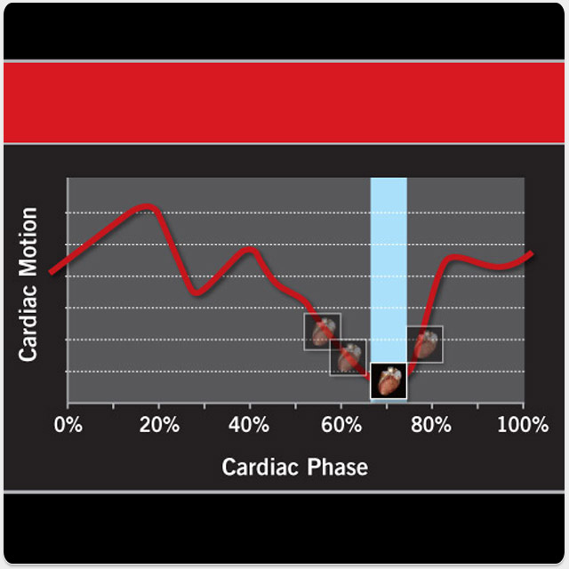
SURECARDIO WITH PHASEXACT
Delivers clear, accurate cardiac images and enhances patient throughput and workflow by automatically selecting optimum scan parameters.
- Automated, clinically validated protocols provide the best temporal resolution regardless of patient heart rate or condition
- Achieve consistent Cardiac CTA with adaptive, multi-segment reconstruction
- Decrease patient scan time through protocol automation
- Reduce view time and storage space with phaseXact, which automatically selects the cardiac phase with the least motion
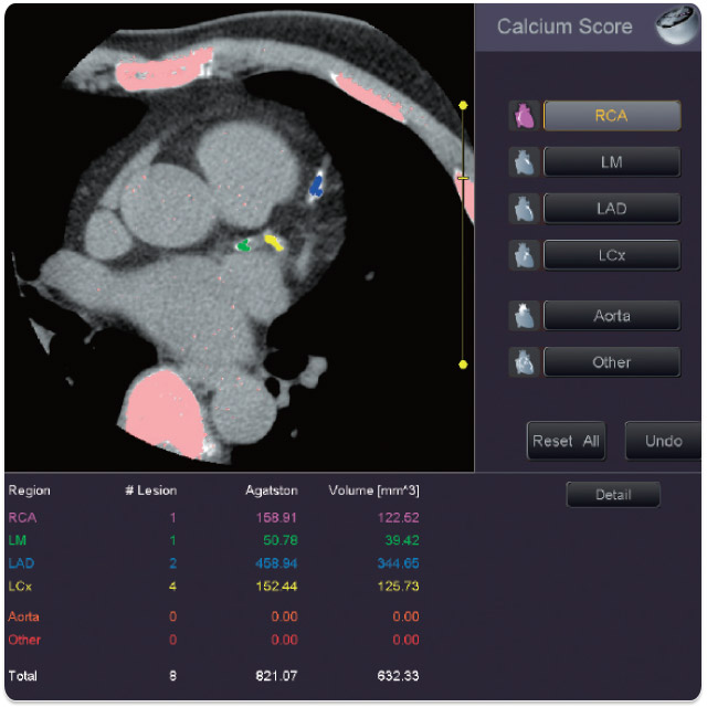
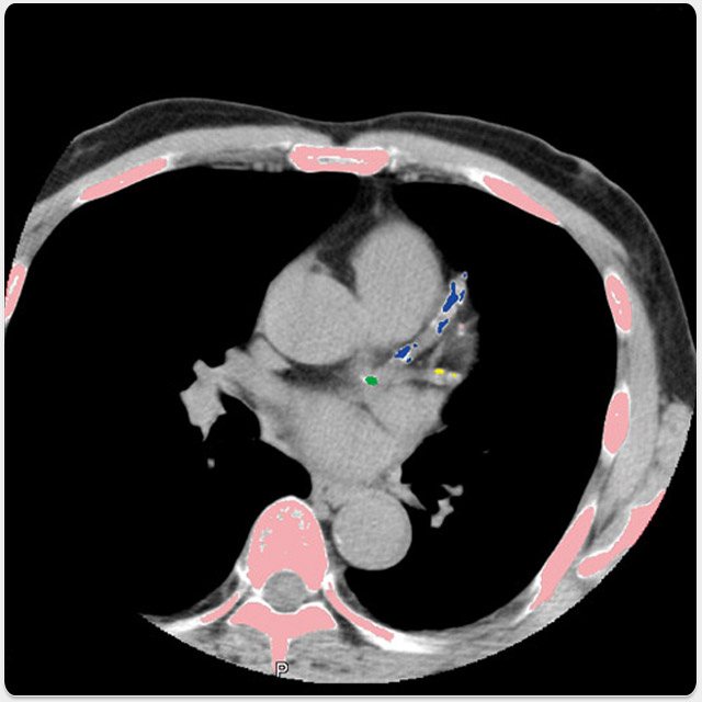
SURECARDIO SCORING
Provides fast and easy evaluation of calcium based on non-contrasted, ECG-gated data directly from the Aquilion console.
- Calculates Ca scores using the Agaston method and the volume mass method
- A report and the representative images showing calcium are automatically generated

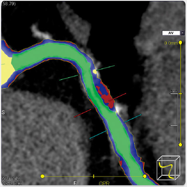
SUREPLAQUE
A comprehensive advanced visualization tool to assist clinicians in evaluating the characteristics inside the blood vessel.
- Visualize coronary vessel anatomy and disease with ease using defined HU ranges
- Quantify plaque burden and coronary remodelling non-invasively
- Characterize lesions in the vessel wall as either calcified or non-calcified
MAXIMISING CLINICAL CAPABILITIES
Aquilion RXL supports Canon’s sophisticated suite of SURETechnologies and other advanced software tools to further increase clinical utility, and incorporates cutting-edge clinical application capabilities to meet customer needs.
CARDIAC
 SURECardio with phaseXact
Accurate cardiac images and enhanced workflow
 SUREPlaque
Automated plaque visualization and characterization
 SURECardio Scoring
Fast and easy evaluation of calcium based on non-contrast ECG-gated data
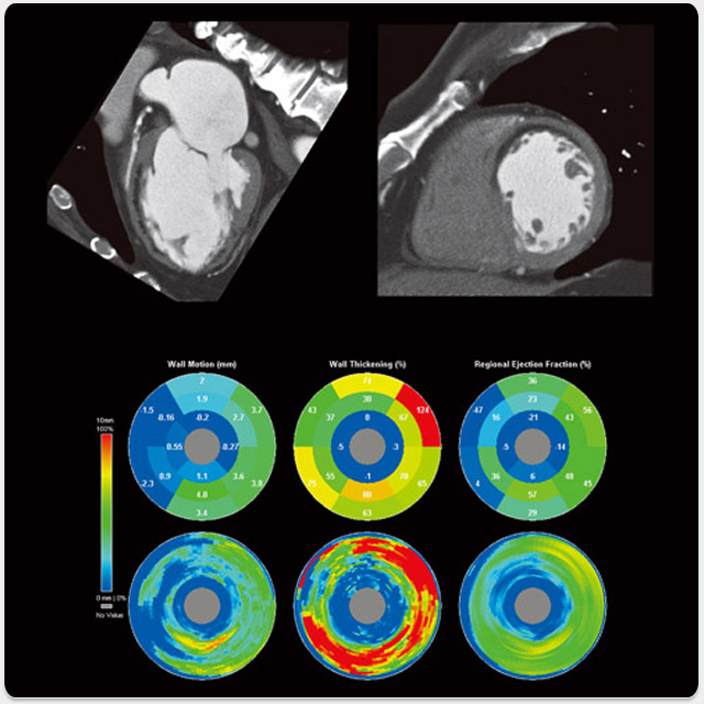 Cardiac Function Analysis
Calculation of various functional parameters, such as ejection fraction, wall motion, and cardiac output
NEURO
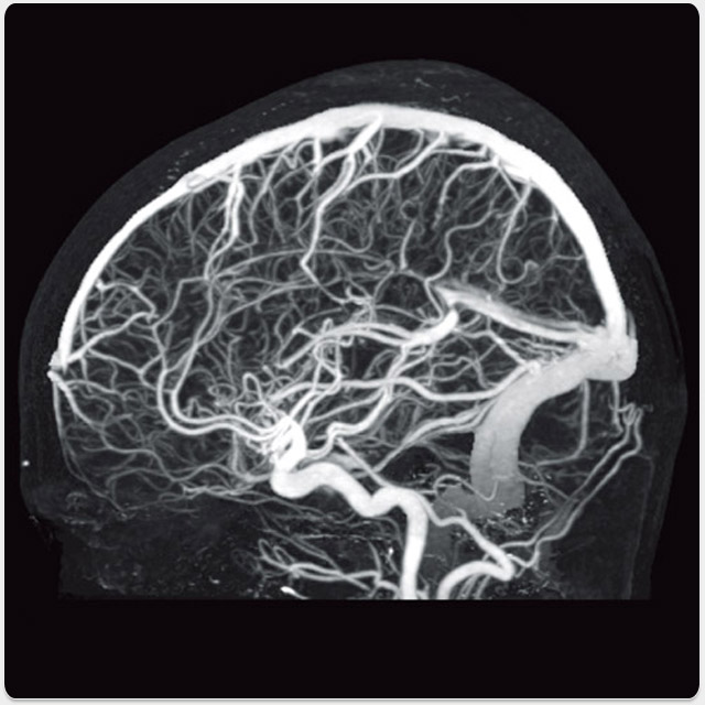 SURESubtraction
Automated digital subtraction of intra-cranial vessels from bone
 CBP Study
Analysis of bloodflow characteristics from dynamic scan images and display of the results as map images
BODY
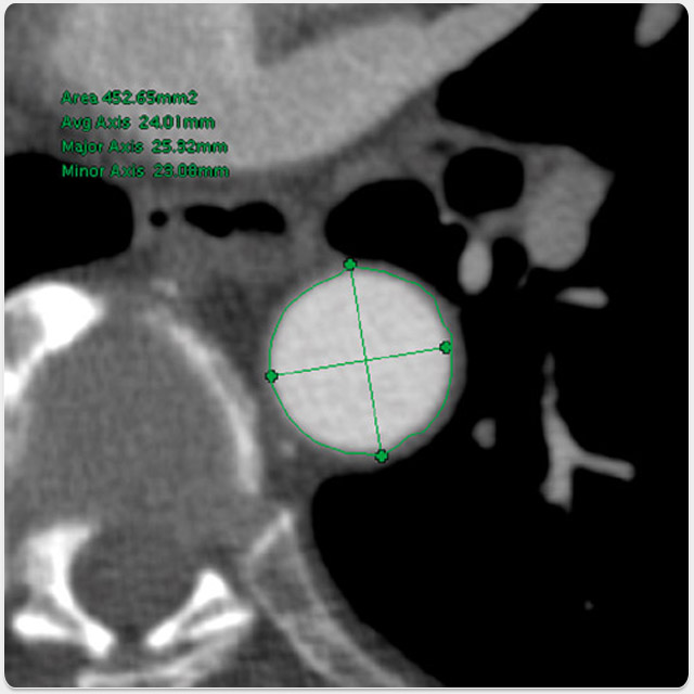 Vessel View
Generation and display of CPR and cross-cut images of blood vessels
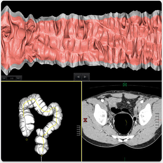 Colon View
Advanced analysis and reporting tools for CT colonoscopy, with display functions such as filet view, fly through, and polyp tagging
 Dental Analysis
Comprehensive dental MPR with easy-to-use tools for pre-operative planning
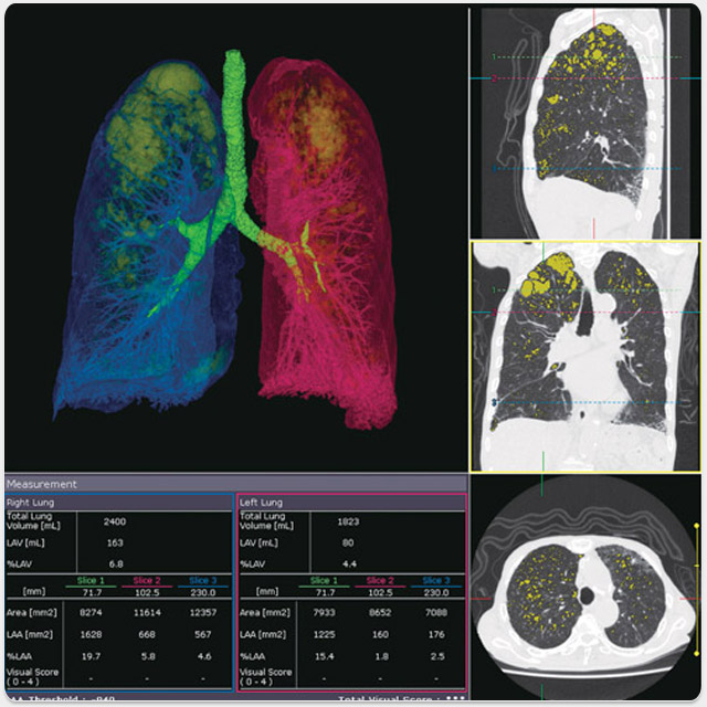 Lung Volume Analysis
Quantification of low attenuation regions in lung tissue (regions of pulmonary emphysema)
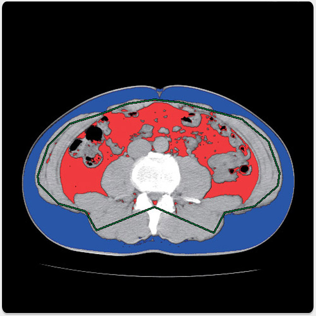 Fat Index View
Automatic calculation of the ratio of visceral to subcutaneous fat as a prognostic indicator of the risk of metabolic syndrome
 SUREFluoro
Realtime reconstruction and display of fluoroscopic images for faster and safer
interventional procedures