Better diagnostics starts here
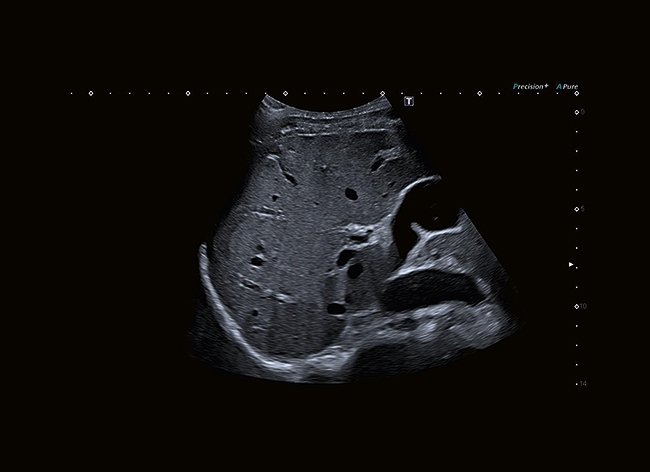
Aplio’s ultra-wideband i-series transducers cover the same bandwidth as two conventional transducers, providing superior sensitivity and resolution for both near and far field. While helping to reduce cost, this revolutionary transducer design can provide better imaging regardless of the patient condition.
Aplio’s Intelligent Dynamic MicroSlice technology increases clinical accuracy and reveals more detail in all depths by electronically sharpening the imaging slice thickness.
![]()
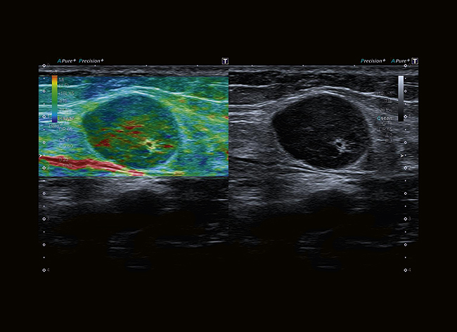
Seeing the unseen with SMI
Experience color flow imaging with unmatched detail and definition on Aplio i800. Superb micro-vascular imaging (SMI) expands the range of visible blood flow to visualize low-velocity microvascular flow never before seen with diagnostic ultrasound.
SMI‘s level of vascular visualization, combined with high frame rates, advances diagnostic confidence when evaluating lesions, cysts and tumors.
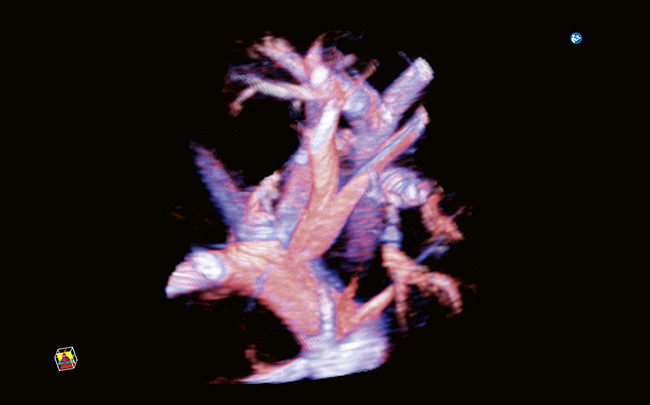
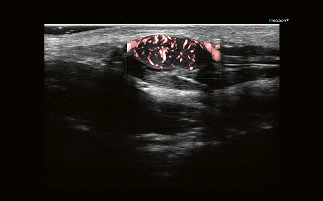
Smart Sensor 3D allows you to acquire accurate 3D volumes with a standard linear or convex transducer, also in SMI mode.
Enjoy the perfect picture

Precision+ offers outstandingly smooth images with sharpened outline of lesions, enhanced image uniformity and reduced clutter.

ApliPure™+ compounding delivers increased imaging contrast and reduced speckle noise to improve visualization.
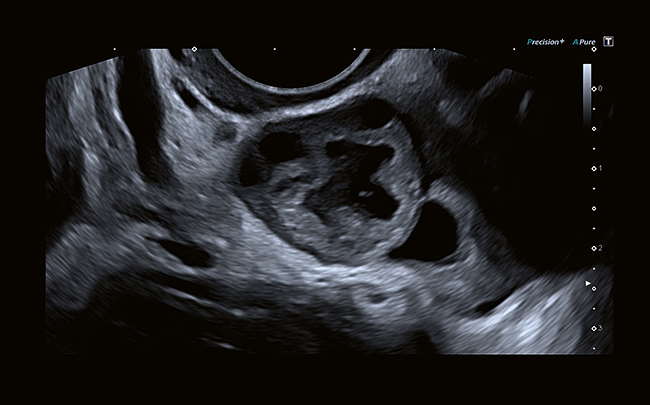
Differential Tissue Harmonics provides harmonic images of unsurpassed spatial resolution, alongside greatly enhanced penetration.

Aplio’s wideband transducer and signal processing technology delivers outstanding sensitivity, penetration and spatial resolution for all Doppler modes.
Amazing detail, outstanding versatility
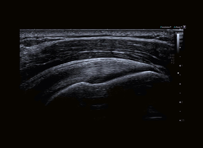
Aplio’s 24 MHz transducer with extended bandwidth and iDMS Dynamic MicroSlice technology provides superior detail and definition in the near field for a wide range of examinations.
 The transducer’s outstanding resolution can help identify fine detail such as layered structures and small lesions.
 Smart 3D is a simple way to add volume-imaging capability to Aplio’s convex and linear transducers, supporting all modes including SMI or shear wave imaging.
 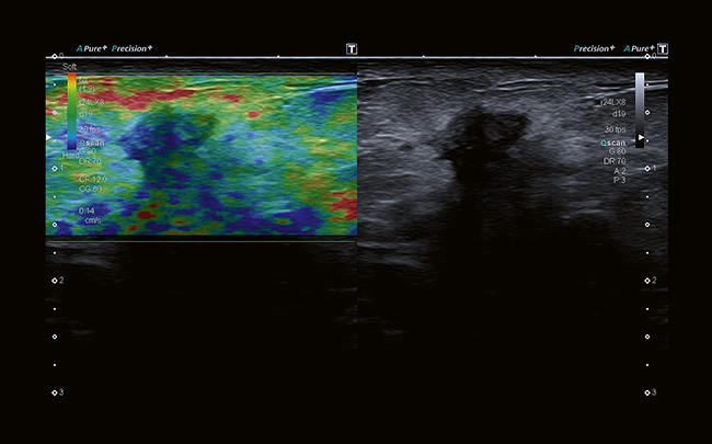 Advanced technologies such as strain elastography or SMI can provide valuable insights into perfusion dynamics or tissue stiffness.