ENJOY THE PERFECT PICTURE EVERYDAY
At Canon we believe that only the best image quality allows a diagnosis to happen quickly and with confidence. Each of our unique imaging technologies provides you with better image quality by reducing noise, strengthening signal and improving visualization. Aplio’s revolutionary High Density Beamformer uses the most advanced digital signal processing to control the ultrasonic beams more precisely and flexibly than any other system.

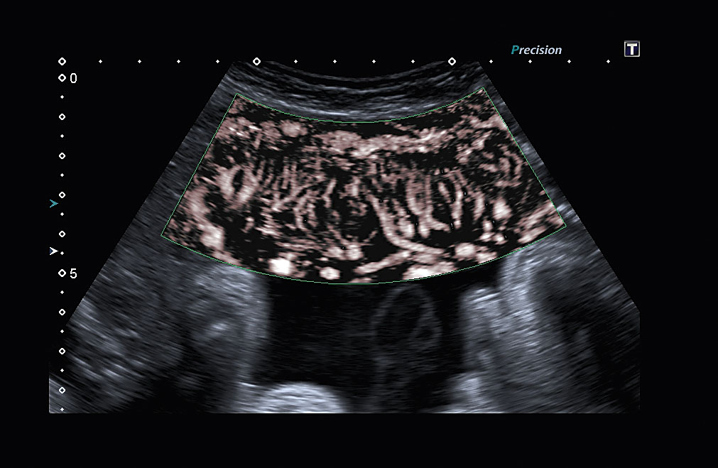
Precision Imaging and Precision+
With Aplio’s new and enhanced Precision Imaging technology you can experience ultrasound imaging as close to reality as never before.

ApliPure™+
ApliPure+ delivers increased imaging contrast and reduced speckle noise to improve visualization.
 Differential Tissue Harmonics
Differential Tissue Harmonic Imaging takes outstanding tissue definition deeper than ever before.

Advanced Dynamic Flow™ (ADF)
Display flow directionally and accurately at high frame rates, while maintaining the full B-mode image quality.
SEEING THE UNSEEN
Canon’s innovative Superb Micro-Vascular Imaging (SMI) technology expands the range of visible blood flow and provides visualization of low velocity microvascular flow seen with ultrasound. SMI’s level of vascular visualization, combined with high frame rates, advances diagnostic confidence when evaluating lesions, cysts and tumors, improving patient outcomes and experience.
Monochrome mode
By removing anatomical background information, the monochrome mode reveals the finest vasculature with high sensitivity.
Color mode
Color-coded SMI demonstrates flow and greyscale information with high temporal and spatial resolution simultaneously.


The principle behind SMI
Traditional color Doppler imaging (left) removes clutter from the images by suppressing low velocity components, resulting in a loss of flow in tiny vessels. SMI (left) separates flow from overlaying tissue motion effectively, while preserving even the subtlest low-flow components with unmatched detail and definition.
A NEW DIMENSION OF IMAGING AND INTERVENTION
Aplio’s comprehensive 3D/4D volume imaging suite extends your diagnostic capabilities into the next dimension of imaging and intervention by providing accurate renderings and arbitrary volume cuts in realtime or offline. Aplio’s new High Density Volume Rendering Engine gives you extraordinary image quality at high volume rates for uncompromised workflow and clinical result.
 Luminance
Luminance is an innovative new surface rendering technique that provides a softer, more natural visualization of the human skin resulting in images of almost photographic impression and quality. The function’s freely movable light source gives you strong visual feedback on depth and detail. Changing the position of the light can help you identify pathological changes and skin defects better and with greater clarity.
 Multi-Planar Reconstruction (MPR)
Aplio’s MPR function allows you to review a specific structure or region of interest simultaneously in three orthogonal planes accompanied by a surface rendering or box volume image. The increased anatomical information contained in the high-resolution cross sections can help you to better understand anatomical relationships or the extent of a given lesion.
 MultiView
With MultiView you can generate series of cross sections of a given volume in an instant. The resulting display of multiple parallel cut planes provides a very effective tool for the assessment of lesions and their associated structures. Multi- View allows you to cut a given volume in any direction to reveal high-resolution off-axis views that can further enhance your diagnostic confidence.


Fly Thru
Fly Thru is a stunning technology that lets you virtually dive into a volume data set to explore cavities, ducts and vessels from the inside and in 3D. Being comparable to virtual endoscopy, Fly Thru adds cross-sectional ultrasound information to the plain surface data, making it an expert tool for exploring lesions and ingrowing masses, as well as to assist in planning and follow-up of interventions such as placing stents or grafts.
A complete range of volume imaging transducers is available for Aplio. Their compact and lightweight design delivers outstanding image quality in an ergonomic housing.